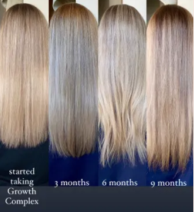
Clinical Studies

VITAMIN C

ABSORBIC ACID CAN REDUCE THE SIGNS OF AGING

Kwak JY, et al. Ascorbyl coumarates as multifunctional cosmeceutical agents that inhibit melanogenesis and enhance collagen synthesis. Arch Dermatol Res. 2015;307(7):635-43.


ABSTRACT

L-Ascorbic acid (AA) and p-coumaric acid (p-CA) are naturally occurring antioxidants that are known to enhance collagen synthesis and inhibit melanin synthesis, respectively. The purpose of this study was to examine hybrid compounds between AA and p-CA as multifunctional cosmeceutical agents. Ascorbyl 3-p-coumarate (A-3-p-C), ascorbyl 2-p-coumarate (A-2-p-C), and their parent compounds were tested for their effects on cellular melanin synthesis and collagen synthesis. At 100 μM, A-3-p-C and A-2-p-C decreased melanin content of human dermal melanocytes stimulated by L-tyrosine, by 65 and 59%, respectively, compared to 11% inhibition of AA and 70% inhibition of p-CA. A-3-p-C and A-2-p-C were less effective than p-CA but more effective than AA at inhibiting tyrosinase activity. A-3-p-C and A-2-p-C were more effective than p-CA at inhibiting the autoxidation of L-3,4-dihydroxyphenylalanine. At 100-300 μM, A-3-p-C and A-2-p-C augmented collagen release from human dermal fibroblasts by 120-144% and 125-191%, respectively, compared to 126-133% increase of AA and 120-146% increase of p-CA. They increased procollagen type I C-peptide release (A-3-p-C, and A-2-p-C) like AA, and decreased matrix metalloproteinase 1 level (A-2-p-C) like p-CA, implicating that they might regulate collagen metabolism by multiple mechanisms. This study suggests that A-3-p-C and A-2-p-C could be used as multifunctional cosmeceutical agents for the attenuation of certain aspects of skin aging.

https://www.ncbi.nlm.nih.gov/pubmed/26078014

VITAMIN C MAY HAVE A PROTECTIVE EFFECT AGAINST CUTANEOUS MELANOMA

Malavolti M, et al. Association between dietary Vitamin C and risk of cutaneous melanoma in a population of Northern Italy. Int J Vitam Nutr Res. 2013;83(5):291-8.


ABSTRACT

Cutaneous melanoma incidence has been increasing during the last few years, and diet has been suggested as one of the lifestyle factors responsible for this increase. Since antioxidant nutrients such as ascorbic acid might prevent skin carcinogenesis, we investigated the risk of cutaneous melanoma related to vitamin C intake in a population-based case-control study in Northern Italy based on 380 melanoma patients and 719 matched controls, to whom we administered a semiquantitative food-frequency questionnaire. After adjusting for potential confounders, odds ratio of melanoma were 0.86 (95 % confidence interval 0.65 – 1.15) and 0.59 (95 % confidence interval 0.37 – 0.94) in the intermediate and highest categories of vitamin C dietary intake respectively, compared with the bottom one. The association between vitamin C and decreased risk persisted after adjustment for some potential confounders. In age- and gender-stratified analyses, this association was seen in young females (< 60 years old), and was found to be enhanced in subjects with phototypes II and III. These results suggest a possible protective activity of vitamin C intake against cutaneous melanoma in specific subgroups of this population of Northern Italy.

http://www.ncbi.nlm.nih.gov/ pubmed/25305224

VITAMIN C ACTS A PHOTO PROTECTANT AND CAN PROTECT THE SKIN FROM UV DAMAGE 

Darr D, et al. Topical Vitamin C protects porcine skin from ultraviolet radiation-induced damage. British Journal of Dermatology. 1992;127(3):247-253.


ABSTRACT

Summary Ultraviolet radiation damage to the skin is due, in part, to the generation of reactive oxygen species. Vitamin C (l‐ascorbic acid) functions as a biological co‐factor and antioxidant due to its reducing properties. Topical application of vitamin C has been shown to elevate significantly cutaneous levels of this vitamin in pigs, and this correlates with protection of the skin from UVB damage as measured by erythema and sunburn cell formation. This protection is biological and due to the reducing properties of the molecule. Further, we provide evidence that the vitamin C levels of the skin can be severely depleted after UV irradiation, which would lower this organ’s innate protective mechanism as well as leaving it at risk of impaired healing after photoinduced damage. In addition, vitamin C protects porcine skin from UVA‐mediated phototoxic reactions (PUVA) and therefore shows promise as a broad‐spectrum photo protectant.

http://onlinelibrary.wiley.com/doi/10.1111/j.1365-2133.1992.tb00122.x/full

COMBINED VITAMIN C AND VITAMIN E REDUCES THE SUNBURN REACTION AND MAY LEAD TO A REDUCED RISK FOR UV-INDUCED SKIN DAMAGE

Eberlein-Konig, B, et al. Protective effect against sunburn of combined systemic ascorbic acid (Vitamin C) and d-alpha-tocopherol (Vitamin E). Journal of the American Academy of Dermatology. 1998;38(1):45-48.


ABSTRACT

Background: UV radiation causes acute adverse effects like sunburn, photosensitivity reactions, or immunologic suppression, as well as long-term sequelae like photoaging or malignant skin tumors. UV radiation induces tissues to produce reactive oxygen species, eicosanoids and cytokines. Inhibition of these mediators might reduce skin damage. Antioxidants such as ascorbic acid and d-α-tocopherol have been found to be photoprotective in some in vitro studies and animal experiments. Objective: Our purpose was to assess the protective effect of systemic vitamins C and E against sunburn in human beings. Methods: In a double-blind placebo-controlled study, each of 10 subjects took daily either 2 gm of ascorbic acid (vitamin C) combined with 1000 IU of d-α-tocopherol (vitamin E) or placebo. The sunburn reaction before and after 8 days of treatment was assessed by determination of the threshold UV dose for eliciting sunburn (minimal erythema dose [MED]) and by measuring the cutaneous blood flow of skin irradiated with incremental UV doses against that of nonirradiated skin. Results: The median MED of those taking vitamins increased from 80 to 96.5 mJ/cm2 (p < 0.01), whereas it declined from 80 to 68.5 mJ/cm2 in the placebo group. Cutaneous blood flow changed significantly (p < 0.05) for most irradiation doses with decreases in those given vitamins and increases in the placebo group. Conclusion: Combined vitamins C and E reduce the sunburn reaction, which might indicate a consequent reduced risk for later sequelae of UV-induced skin damage. The increase of sunburn reactivity in the placebo group could be related to “priming” by the previous UV exposure. (J Am Acad Dermatol 1998;38:45-8.)

www.sciencedirect.com/science/article/pii/S0190962298705377

VITAMIN C AND E IN A COMBINATION MAY PROTECT AGAINST SKIN CANCER AND PHOTOAGING

Lin JY, et al. UV photoprotection by combination topical antioxidants Vitamin C and Vitamin E. Journal of the American Academy of Dermatology. 2003;48(6):866-874.


ABSTRACT

Background: Virtually all plants and animals protect themselves from the sun using vitamins C and E. Objective: The purpose of this study was to see if a combination of topical vitamins C and E is better for UV protection to skin than an equivalent concentration of topical vitamin C or E alone. Methods: We developed a stable aqueous solution of 15% L-ascorbic acid (vitamin C) and 1% α-tocopherol (vitamin E). We applied antioxidant or vehicle solutions to pig skin daily for 4 days. We irradiated (1-5× minimal erythema dose) control- and antioxidant-treated skin using a solar simulator with a 295-nm band-pass filter. On day 5, we measured antioxidant protection factor, erythema, sunburn cells, and thymine dimers. Results: The combination of 15% L-ascorbic acid and 1% α-tocopherol provided significant protection against erythema and sunburn cell formation; either L-ascorbic acid or 1% α-tocopherol alone also was protective but the combination was superior. Application during 4 days provided progressive protection that yielded an antioxidant protection factor of 4-fold. In addition, the combination of vitamins C and E provided protection against thymine dimer formation. Conclusion: Appreciable photoprotection can be obtained from the combination of topical vitamins C and E. We suggest that these natural products may protect against skin cancer and photoaging. (J Am Acad Dermatol 2003;48:866-74.) www.sciencedirect.com/science/article/pii/S0190962203007813

VITAMIN C CAN EXTEND CELLULAR VIABILITY OF SKIN CELLS AND PROMOTES THE FORMULATION OF THE EPIDERMAL BARRIER

Boyce ST, et al. Vitamin C regulates keratinocyte viability, epidermal barrier, and basement membrane in vitro, and reduces wound contraction after grafting of cultured skin substitutes. Journal of Investigative Dermatology. 2002;118(4):565-572.


ABSTRACT

Cultured skin substitutes have become useful as adjunctive treatments for excised, full-thickness burns, but no skin substitutes have the anatomy and physiology of native skin. Hypothetically, deficiencies of structure and function may result, in part, from nutritional deficiencies in culture media. To address this hypothesis, vitamin C was titrated at 0.0, 0.01, 0.1, and 1.0 mM in a cultured skin substitute model on filter inserts. Cultured skin substitute inserts were evaluated at 2 and 5 wk for viability by incorporation of 5-bromo-2′-deoxyuridine (BrdU) and by 3-(4,5-dimethylthiazol-2-yl)-2,5-diphenyl tetrazolium bromide (MTT) conversion. Subsequently, cultured skin substitute grafts consisting of cultured human keratinocytes and fibroblasts attached to collagen-glycosaminoglycan substrates were incubated for 5 wk in media containing 0.0 mM or 0.1 mM vitamin C, and then grafted to athymic mice. Cultured skin substitutes (n = 3 per group) were evaluated in vitro at 2 wk of incubation for collagen IV, collagen VII, and laminin 5, and through 5 wk for epidermal barrier by surface electrical capacitance. Cultured skin substitutes were grafted to full-thickness wounds in athymic mice (n = 8 per group), evaluated for surface electrical capacitance through 6 wk, and scored for percentage original wound area through 8 wk and for HLA-ABC-positive wounds at 8 wk after grafting. The data show that incubation of cultured skin substitutes in medium containing vitamin C results in greater viability (higher BrdU and MTT), more complete basement membrane development at 2 wk, and better epidermal barrier (lower surface electrical capacitance) at 5 wk in vitro. After grafting, cultured skin substitutes with vitamin C developed functional epidermal barrier earlier, had less wound contraction, and had more HLA-positive wounds at 8 wk than without vitamin C. These results suggest that incubation of cultured skin substitutes in medium containing vitamin C extends cellular viability, promotes formation of epidermal barrier in vitro, and promotes engraftment. Improved anatomy and physiology of cultured skin substitutes that result from nutritional factors in culture media may be expected to improve efficacy in treatment of full-thickness skin wounds.

www.sciencedirect.com/science/article/pii/S0022202X15416100


VITAMIN E

VITAMIN E REDUCES ACUTE AND CHRONIC SKIN DAMAGE INDUCED BY UV RADIATION. IT ALSO REDUCES INFLAMMATION AND PIGMENTATION AND REDUCES RISK OF SKIN CANCER.

Burke KE, et al. Effects of topical and oral vitamin E on pigmentation and skin cancer induced by ultraviolet irradiation in Skh:2 hairless mice. Nutr Cancer. 2000;38(1);87-97.


ABSTRACT

This study investigates whether supplementation with topical RRR-alpha-tocopherol (Eol), topical RRR-alpha-tocopheryl succinate, and oral RRR-alpha-tocopheryl acetate can reduce the incidence of acute and chronic damage to the skin (i.e., sunburn and pigmentation and skin cancer, respectively) induced by ultraviolet (UV) irradiation to mice. Groups of twenty Skh:2 female hairless pigmented mice were treated with 1) lotion vehicle, 2) 5% Eol lotion, 3) 5% topical RRR-alpha-tocopheryl succinate lotion, or 4) lotion vehicle and oral RRR-alpha-tocopheryl acetate. Within each group, 15 mice were exposed to 0.24 J/cm2 of UV-B radiation three times per week. The animals’ weights and food intakes were monitored, and the vitamin E concentrations of skin, liver, and adipose tissue were measured to determine whether the topical Eol resulted in significant tissue levels. Skin pigmentation was scored, and the total number of clinically detectable skin tumors per animal was counted weekly. Results showed that the skin concentrations of Eol, as well as levels in the adipose tissue, were increased after topical application. Mice treated with each form of vitamin E showed no signs of toxicity and had significantly less acute and chronic skin damage induced by UV irradiation, as indicated by reduced inflammation and pigmentation and by later onset and lesser incidence of skin cancer.

https://www.ncbi.nlm.nih.gov/pubmed/11341050

YELLOW NAIL SYNDROME IMPROVED WITH TOPICAL VITAMIN E TO THE NAIL PLATES AND PERIUNGUAL SKIN

Lambert EM, et al. Yellow nail syndrome in three siblings: A randomized double-blind trial of topical Vitamin E. Pediatric Dermatology. 2006;23(4):390-395.


ABSTRACT

Abstract:  Yellow nail syndrome is an uncommon disorder characterized by dystrophic nails, lymphedema, and respiratory disease. It has rarely been reported in children and this is the first report of congenital yellow nails in siblings. The purpose of this study was to determine whether topical vitamin E applied to the nail plates and periungual skin would affect the growth rate or appearance of the fingernails in patients with congenital yellow nail syndrome. This study was the first trial of a treatment for this entity in children and the largest randomized double blind trial to date. We found that vitamin E solution had no significant effect (p = 0.84) on fingernail growth or the global appearance score (p = 1.0) when compared with placebo. The average growth rates and global assessment scores improved and onycholysis and onychomadesis decreased from baseline with both vitamin E and placebo treatment, although these were not primary end points of the study. Topical vitamin E did not result in a statistically significant improvement when compared with oil alone for the treatment of the nails in our three patients with yellow nail syndrome. However, it is interesting and perhaps clinically useful that both vitamin E and placebo oil improved the condition of the nails.

http://onlinelibrary.wiley.com/doi/10.1111/j.1525-1470.2006.00251.x/full

VITAMIN C AND E COMBINATION SIGNIFICANTLY REDUCES THE UVB-INDUCED DNA DAMAGE IN SKIN, SUGGESTING THAT IT IS AN ANTIOXIDANT TREATMENT TO PROTECT AGAINST DNA DAMAGE

Placzek M, et al. Ultraviolet B-induced DNA damage in human epidermis is modified by the antioxidants ascorbic acid and D-alpha-tocopherol. J Invest Dermatol. 2005;124(2):304-7.


ABSTRACT

DNA damage caused by ultraviolet (UV) irradiation is considered the main etiologic factor contributing to the development of skin cancer. Systemic or topical application of antioxidants has been suggested as a protective measure against UV-induced skin damage. We investigated the effect of long-term oral administration of a combination of the antioxidants ascorbic acid (vitamin C) and D-alpha-tocopherol (vitamin E) in human volunteers on UVB-induced epidermal damage. The intake of vitamins C and E for a period of 3 mo significantly reduced the sunburn reaction to UVB irradiation. Detection of thymine dimers in the skin using a specific antibody revealed a significant increase of this type of DNA damage following UVB exposure. After 3 mo of antioxidant administration, significantly less thymine dimers were induced by the UVB challenge, suggesting that antioxidant treatment protected against DNA damage.

https://www.ncbi.nlm.nih.gov/pubmed/15675947

VITAMIN C AND VITAMIN E COMBINATION REDUCES SUNBURN REACTIONS AND REDUCES RISK FOR UV INDUCED SKIN DAMAGE

Eberlein-Konig B, et al. Protective effect against sunburn of combined systemic ascorbic acid and d-alpha-tocopherol (Vitamin E). J Am Acad Dermatol. 1998;38(1):45-8.


ABSTRACT

BACKGROUND: UV radiation causes acute adverse effects like sunburn, photosensitivity reactions, or immunologic suppression, as well as long-term sequelae like photoaging or malignant skin tumors. UV radiation induces tissues to produce reactive oxygen species, eicosanoids and cytokines. Inhibition of these mediators might reduce skin damage. Antioxidants such as ascorbic acid and d-alpha-tocopherol have been found to be photoprotective in some in vitro studies and animal experiments.

OBJECTIVE: Our purpose was to assess the protective effect of systemic vitamins C and E against sunburn in human beings.

METHODS: In a double-blind placebo-controlled study, each of 10 subjects took daily either 2 gm of ascorbic acid (vitamin C) combined with 1000 IU of d-alpha-tocopherol (vitamin E) or placebo. The sunburn reaction before and after 8 days of treatment was assessed by determination of the threshold UV dose for eliciting sunburn (minimal erythema dose [MED]) and by measuring the cutaneous blood flow of skin irradiated with incremental UV doses against that of non irradiated skin.

RESULTS: The median MED of those taking vitamins increased from 80 to 96.5 mJ/cm2 (p < 0.01), whereas it declined from 80 to 68.5 mJ/cm2 in the placebo group. Cutaneous blood flow changed significantly (p < 0.05) for most irradiation doses with decreases in those given vitamins and increases in the placebo group.

CONCLUSION: Combined vitamins C and E reduce the sunburn reaction, which might indicate a consequent reduced risk for later sequelae of UV-induced skin damage. The increase of sunburn reactivity in the placebo group could be related to “priming” by the previous UV exposure.

https://www.ncbi.nlm.nih.gov/pubmed/9448204


BIOTIN

BIOTIN INCREASES NAIL THICKNESS AND HELPS WITH BRITTLE NAILS

Hochman LG, et al. Brittle nails: response to daily biotin supplementation.

Cutis. 1993;51(4):303-5.


ABSTRACT

A recent study from Switzerland demonstrated a 25 percent increase in nail plate thickness in patients with brittle nails who received biotin supplementation. Analysis of all visits to a nail consultation practice over a six-month period revealed forty-four patients with this condition who had been prescribed the B-complex vitamin biotin. Of these, thirty-five who took daily supplementation were subjectively evaluated. Twenty-two of thirty-five (63 percent) showed clinical improvement and thirteen (37 percent) reported no change in their condition. The results of this small, retrospective study suggest a positive response to biotin in the treatment of brittle nails in some patients.

https://www.ncbi.nlm.nih.gov/pubmed/8477615

ORAL BIOTIN  SHOWED SIGNIFICANT IMPROVEMENTS FOR THOSE WITH UNCOMBABLE HAIR SYNDROME, WITH AN INCREASE IN AHIR GROWTH RATE, STRENGTH AND COMBABILITY OF HAIR.

Shelley WB, et al. Uncombable hair syndrome: observations on response to biotin and occurrence in siblings with ectodermal dysplasia. J Am Acad Dermatol. 1985;13(1):97-102.


ABSTRACT

Three children are reported with uncombable hair syndrome, consisting of slow-growing, straw-colored scalp hair that could not be combed flat. The hairs appeared normal on light microscopy but on scanning electron microscopy were triangular in cross section, with canal-like longitudinal depressions. Oral biotin, 0.3 mg three times a day, produced significant improvement after 4 months in one patient, with increased growth rate and with strength and combability of the hair, although the triangular shape remained. The other two patients were unique in having associated ectodermal dysplasia. Their hair slowly improved in appearance and combability over 5 years without biotin therapy.

https://www.ncbi.nlm.nih.gov/pubmed/4031156

BIOTIN SUPPLEMENTATION MAY BENEFIT AILMENTS, SUCH AS BRITTLE HAIR SYNDROME OR UNCOMBABLE HAIR

Patel DP, et al. A review of the use of biotin for hair loss. Skin Appendage Disord. 2017;3(3):166-169.


ABSTRACT

BACKGROUND: Biotin has gained commercial popularity for its claimed benefits on healthy hair and nail growth. Despite its reputation, there is limited research to support the utility of biotin in healthy individuals.

OBJECTIVE: To systematically review the literature on biotin efficacy in hair and nail growth.

METHODS: We conducted a PubMed search of all case reports and randomized clinical trials (RCTs) using the following terms: (biotin and hair); (biotin and supplementation and hair); (biotin supplementation); (biotin and alopecia); (biotin and nails); (biotin and dermatology), and (biotin recommendations).

RESULTS: We found 18 reported cases of biotin use for hair and nail changes. In all cases, patients receiving biotin supplementation had an underlying pathology for poor hair or nail growth. All cases showed evidence of clinical improvement after receiving biotin.

CONCLUSIONS: Though its use as a hair and nail growth supplement is prevalent, research demonstrating the efficacy of biotin is limited. In cases of acquired and inherited causes of biotin deficiency as well as pathologies, such as brittle nail syndrome or uncombable hair, biotin supplementation may be of benefit. However, we propose these cases are uncommon and that there is lack of sufficient evidence for supplementation in healthy individuals.

https://www.ncbi.nlm.nih.gov/pubmed/28879195

BIOTIN SUPPLEMENTATION IMPROVED HAIR APPEARANCE IN INDIVIDUALS WITH  UNCOMBABLE HAIR AND ALSO IMPROVED NAIL FRAGILITY

Boccaletti V, et al. Familial uncombable hair syndrome: ultrastructural hair study and response to biotin. Pediatric Dermatology. 2007;24(3):E14-E16.


ABSTRACT

We report a family affected to the fourth generation by uncombable hair syndrome. This syndrome is characterized by unruly, dry, blond hair with a tangled appearance. The family pedigree strongly supports the hypothesis of autosomal dominant inheritance; some members of the family had, apart from uncombable hair, minor signs of atopy and ectodermal dysplasia, such as abnormalities of the nails. The diagnosis was confirmed by means of extensive scanning electron microscopy. A trial with oral biotin 5mg/day was started on two young patients with excellent results as regards the hair appearance, although scanning electron microscopy did not show structural changes in the hair. After a 2‐year‐period of follow‐up, hair normality was maintained without biotin, while nail fragility still required biotin supplementation for control.

http://onlinelibrary.wiley.com/doi/10.1111/j.1525-1470.2007.00385.x/abstract


D-CALCIUM PANTOTHENATE

PANTHOTHENIC ACID AND ASCORBIC ACID MAY REDUCE THE SIGNS OF SKIN SCARRING

Vaxman F, et al. Effect of pantothenic acid and ascorbic acid supplementation on human skin wound healing process. European Surgical Research. 1995;27:158-166.


ABSTRACT

This study aimed at testing human skin wound healing improvement by a 21-day supplementation of 1.0 g ascorbic acid (AA) and 0.2 g pantothenic acid (PA). 49 patients undergoing surgery for tattoos, by the successive resections procedure, entered a double-blind, prospective and randomized study. Tests performed on both skin and scars determined: hydroxyproline concentrations, number of fibroblasts, trace element contents and mechanical properties. In the 18 supplemented patients, it was shown that in skin (day 8) Fe increased (p < 0.05) and Mn decreased (p < 0.05); in scars (day 21), Cu (p = 0.07) and Mn (p < 0.01) decreased, and Mg (p < 0.05) increased; the mechanical properties of scars in group A were significantly correlated to their contents in Fe, Cu and Zn, whereas no correlation was shown in group B. In blood, AA increased after surgery with supplementation, whereas it decreased in controls. Although no major improvement of the wound healing process could be documented in this study, our results suggest that the benefit of AA and PA supplementation could be due to the variations of the trace elements, as they are correlated to mechanical properties of the scars.

https://www.karger.com/Article/Abstract/129395

PANTHOTHENIC ACID SIGNIFICANTLY REDUCES INFLAMMATORY BLEMISHES AND LESIONS CAUSED BY ACNE

Yang M, et al. A randomized, double-blind, placebo-controlled study of a novel pantothenic acid-based dietary supplement in subjects with mild to moderate facial acne. Dermatol Ther. 2014;4(1):93-101.


ABSTRACT

INTRODUCTION: The purpose of this study was to determine the safety, tolerability and effectiveness of daily administration of an orally administered pantothenic acid-based dietary supplement in men and women with facial acne lesions.

METHODS: A randomized, double-blind, placebo-controlled study of adults previously diagnosed with mild to moderate acne vulgaris was performed. Subjects were randomized to the study agent, a pantothenic acid-based dietary supplement, or a placebo for 12 weeks (endpoint). The primary outcome of the study was the difference in total lesion count between the study agent group versus the placebo group from baseline to endpoint. Secondary measurements included differences in mean non-inflammatory and inflammatory lesions, Investigators Global Assessment and Dermatology Life Quality Index (DLQI) scores between the two groups. Investigator assessment of overall improvement and skin photographs were also taken. Safety and tolerability endpoints were the assessment of adverse events and measurement of serum complete blood count and hepatic function.

RESULTS: Forty-eight subjects were enrolled and 41 were evaluable. There was a significant mean reduction in total lesion count in the pantothenic acid group versus placebo at week 12 (P = 0.0197). Mean reduction in inflammatory lesions was also significantly reduced and DLQI scores were significantly lower at week 12 in the pantothenic acid group versus placebo. The study agent was safe and well tolerated.

CONCLUSIONS: The results from this study indicate that the administration of a pantothenic acid-based dietary supplement in healthy adults with facial acne lesions is safe, well tolerated and reduced total facial lesion count versus placebo after 12 weeks of administration. Secondary analysis shows that the study agent significantly reduced area-specific and inflammatory blemishes. Further randomized, placebo-controlled trials are warranted.

http://www.ncbi.nlm.nih.gov/ pubmed/24831048


ZINC

ZINC SUPPLEMENTATION REVERSED SIGNIFICANT HAIR LOSS

Neve HJ, et al. Reversal of hair loss following vertical gastroplasty when treated with zinc sulphate. Obesity Surgery. 1996;6(1):63-65.


ABSTRACT

Background: Nutritional complications following surgery for morbid obesity include both vitamin and mineral deficiency. Severe cases of zinc deficiency can lead to alopecia, diarrhoea, emotional disorders, weight loss, intercurrent infection, bullous-pustular dermatitis and hypogonadism in males. Hair loss may occur after vertical gastroplasty (VG). Methods: Diffuse hair loss occurred in 47 out of 130 patients who underwent VG. All patients had been routinely advised to take a multivitamin supplement, but 47 developed hair loss despite taking the supplement. These patients were then prescribed Zinc Sulphate 200 mg three times a day. There was no alteration in the vitamin supplementation. Results: Arrest of hair loss and regrowth occurred in all patients. However, five patients reported recurrence of hair loss after stopping zinc. This loss was reversed within 6 months of recommencing zinc 600 mg daily. Ten control patients had no hair loss after gastrointestinal surgery. Conclusion: Significant hair loss occurred in about one-third of patients after VG, and was reversed by zinc supplementation.

https://link.springer.com/article/10.1381/096089296765557295

ZINC SUPPLEMENTATION SIGNIFICANTLY ACCELERATES THE RE-GROWTH OF HAIR

Plonka PM, et al. Zinc as an ambivalent but potent modulator of murine hair growth in vivo – preliminary observations. Experimental Dermatology. 2005;14(11):844-853.


ABSTRACT

Abstract:  Oral zinc (Zn2+) is often employed for treating hair loss, even in the absence of zinc deficiency, although its mechanisms of action and efficacy are still obscure. In the current study, we explored the in vivo effects of oral zinc using the C57BL/6 mouse model for hair research. Specifically, we investigated whether continuous administration of high‐dose ZnSO4 × 7H2O (20 mg/ml) in drinking water affects hair follicle (HF) cycling, whether it retards or inhibits chemotherapy‐induced alopecia (CIA) and whether it modulates the subsequent hair re‐growth pattern. Here, we show that high doses of oral zinc significantly inhibit hair growth by retardation of anagen development. However, oral zinc also significantly retards and prolongs spontaneous, apoptosis‐driven HF regression (catagen). Oral zinc can also retard, but not prevent, the onset of CIA in mice. Interestingly, Zn2+ treatment of cyclophosphamide‐damaged HFs also significantly accelerates the re‐growth of normally pigmented hair shafts, which reflects a promotion of HF recovery. However, if given for a more extended time period, zinc actually retards hair re‐growth. Thus, high‐dose oral zinc is a powerful, yet ambivalent hair growth modulator in mice, whose ultimate effects on the HF greatly depend on the timing and duration of zinc administration. The current study also encourages one to explore whether oral zinc can mitigate chemotherapy‐induced hair loss in humans and/or can stimulate hair re‐growth.

http://onlinelibrary.wiley.com/doi/10.1111/j.1600-0625.2005.00365.x/full

ZINC SUPPLEMENTATION INCREASED HAIR GROWTH IN PATIENTS WITH ALOPECIA

Park H, et al. The therapeutic effect and the changed serum zinc level after zinc supplementation in alopecia areata patients who had a low serum zinc level. Ann Dermatol. 2009;21(2):142-146.


ABSTRACT

It has been reported that some alopecia areata patients have zinc deficiency. There have also been several reports published concerning oral zinc sulfate therapy, with encouraging results, in some alopecia areata patients. The purpose of this study was to evaluate the therapeutic effects of oral zinc supplementation for twelve weeks in alopecia areata patients who had a low serum zinc level. Oral zinc gluconate (50 mg/T/day) supplementation was given to alopecia areata patients without any other treatment for twelve weeks. The serum zinc level was measured before and after zinc supplementation. A four-point scale of hair regrowth was used to evaluate the therapeutic effect of oral zinc supplementation in these patients. Fifteen alopecia areata patients were enrolled in this study. After the therapy, the serum zinc levels increased significantly from 56.9 microg/ to 84.5 microg/dl. Positive therapeutic effects were observed for 9 out of 15 patients (66.7%) although this was not statistically significant. The serum zinc levels of the positive response group increased more than those of the negative response group (p=0.003). Zinc supplementation needs to be given to the alopecia areata patients who have a low serum zinc level. We suggest that zinc supplementation could become an adjuvant therapy for the alopecia areata patients with a low serum zinc level and for whom the traditional therapeutic methods have been unsuccessful.


The Therapeutic Effect and the Changed… (PDF Download Available). Available from: https://www.researchgate.net/publication/44649764_The_Therapeutic_Effect_and_the_Changed_Serum_Zinc_Level_after_Zinc_Supplementation_in_Alopecia_Areata_Patients_Who_Had_a_Low_Serum_Zinc_Level

LOW ZINC OR DISTURBANCES IN ZINC METABOLISM ARE ASSOCIATED WITH HAIR LOSS

Kil MS, et al. Analysis of serum zinc and copper concentrations in hair loss. Ann Dermatol. 2013;25(4):405-409.


ABSTRACT


Background:It is well known that some trace elements such as zinc and copper play a significant role in many forms of hair loss. However, the effect of zinc and copper in the pathogenesis of hair loss is still unknown.


Objective: The purpose of this study is to evaluate the zinc and copper status in each of four types of hair loss.

Methods: A study was carried out with 30 health controls and 312 patients who were diagnosed with alopecia areata (AA), male pattern hair loss, female pattern hair loss and telogen effluvium (TE) (2008 to 2011; Hallym University Kangdong Sacred Heart Hospital). Zinc and copper serum concentrations were evaluated between controls and each of four types of hair loss patients.

Results: In all of the hair loss patients, the mean serum zinc was 84.33±22.88, significantly lower than the control group (97.94±21.05 µg/dl) (p=0.002), whereas the serum copper was 96.44±22.62, which was not significantly different (p=0.975). The analysis of each group showed that all groups of hair loss had statistically lower zinc concentration, but not copper concentrations. However, the ratio of the patients with serum zinc concentration lower than 70 µg/dl was significantly high in only the AA group (odds ratio, OR 4.02; confidence interval, CI 1.13 to 14.31) and the TE group (OR 1.12; CI 1.12 to 17.68).

Conclusion: The data led to the hypothesis of zinc metabolism disturbances playing a key role in hair loss, especially AA and TE, whereas the effect of copper on hair growth and shedding cycles still needs more study.

https://synapse.koreamed.org/DOIx.php?id=10.5021/ad.2013.25.4.405

ZINC SUPPLEMENTATION SIGNIFICANTLY IMPROVED YELLOW NAIL SYNDROME

Arroyo JF, et al. Improvement of yellow nail syndrome with oral zinc supplementation. Clinical and Experimental Dermatology. 1993.


ABSTRACT


An unusual case of yellow nail syndrome (YNS) is reported. Total resolution of yellow nails and lymphoedema was observed following oral zinc supplementation for 2 years. A few years later, the patient developed a classical seropositive rheumatoid arthritis (RA). YNS, alone or associated with RA remains a rare clinical condition. The reported beneficial effects of zinc supplementation in YNS, as well as in several other pathological conditions, raise interest about the role of this trace element and its potential therapeutic implications and suggest further investigations are necessary.

http://onlinelibrary.wiley.com/doi/10.1111/j.1365-2230.1993.tb00971.x/full


SELENIUM

SELENIUM CAN HELP PREVENT DANDRUFF

Sheth RA. A comparison of miconazole nitrate and selenium disulfide as anti-dandruff agents. International Journal of Dermatology. 1983;22(2):123-125.


ABSTRACT

ABSTRACT: The anti‐dandruff efficacy of two shampoos containing 2% miconazole nitrate and 2.5% selenium disulfide was compared in 15 subjects and eight subjects, respectively. The antifungal drug, miconazole nitrate, was found to possess anti‐dandruff activity similar to selenium sulfide, a cytostatic compound. For evaluation, techniques of clinical grading and the corneocyte count were used and the latter was found inaccurate. Instead, clinical assessment by grading the severity, supplemented by cytodiagnosis by smear examination was found more helpful. The latter helps to evaluate both the altered pathophysiologic and microbial factors operating in a given case of dandruff, which is a symptom complex brought about by multiple etiologic factors.

http://onlinelibrary.wiley.com/doi/10.1111/j.1365-4362.1983.tb03330.x/full

SELENIUM IS AN IMPORTANT COMPONENT OF CELLULAR ANTIOXIDANT DEFENSES AND MAY PROTECT AGAINST UV RADIATION INDUCED DAMAGE TO SKIN CELLS

McKenzie RC. Selenium, ultraviolet radiation and the skin. Clinical and Experimental Dermatology. 2000;25(8):631-636.


ABSTRACT

Selenium (Se) is a dietary trace mineral in which there has recently been a surge of interest, in both the popular and the scientific press, because of its demonstrated anti‐carcinogenic and anti‐inflammatory properties in humans. In this short review, I will explain why Se is an important component of cellular anti‐oxidant defences and review its protective effects against UV radiation‐induced damage to skin cells. Although little is known about whether selenium can protect human skin from UV‐induced damage, clinical studies are underway and the anti‐oxidant may offer considerable benefits.

http://onlinelibrary.wiley.com/doi/10.1046/j.1365-2230.2000.00725.x/full


CYNATINE HNS

CYNATINE HNS IS AN EFFECTIVE SUPPLEMENT FOR IMPROVING SKIN IN 90 DAYS OF LESS.  CYNATINE HNS IMPROVED MOISTURE, SKIN ELASTICITY, WRINKLE REDUCTION, SKIN COMPACTNESS AND SKIN APPEARANCE

Beer C, et al. A randomized, double-blind, placebo-controlled clinical trial to investigate the effect of Cynatine HNS on skin characteristics. Int J Cosmet Sci. 2013;35(6):608-12.


ABSTRACT

OBJECTIVE: A new, novel product, Cynatine(®) HNS was evaluated for its effects as a supplement for improving various aspects of skin in a randomized, double-blind, placebo-controlled clinical trial.

METHODS: A total of 50 females were included and randomized into two groups. The active group (n = 25) received two capsules totalling of Cynatine(®) HNS, comprised of Cynatine(®) brand keratin (500 mg) plus vitamins and minerals, per day, and the placebo group (n = 25) received two identical capsules of maltodextrin per day for 90 days. End points for skin moisture, skin elasticity, wrinkle reduction, skin compactness and skin appearance were measured.

RESULTS: The results show that subjects taking Cynatine(®) HNS showed statistically significant improvements in their skin when compared with placebo.

CONCLUSION: Cynatine(®) HNS is an effective supplement for improving skin in 90 days or less


https://www.ncbi.nlm.nih.gov/pubmed/23902431

CYNATINE HNS IMPROVED HAIR GROWTH, AMINO ACID COMPOSITION AND HAIR LUSTER. NAIL STRENGTH AND APPEARANCE WAS ALSO IMPROVED AFTER 90 DAYS

Beer C, et al. A clinical trial to investigate the effect of Cynatine HNS on hair and nail parameters. Scientific World Journal. 2014.


ABSTRACT

OBJECTIVE: A new, novel product, Cynatine HNS, was evaluated for its effects as a supplement for improving various aspects of hair and nails in a randomized, double-blind, placebo-controlled clinical trial.

METHODS: A total of 50 females were included and randomized into two groups. The active group (n = 25) received 2 capsules containing Cynatine HNS, comprised of Cynatine brand keratin (500 mg) plus vitamins and minerals, per day, and the placebo group (n = 25) received 2 identical capsules of maltodextrin per day for 90 days. End points for hair loss, hair growth, hair strength, amino acid composition, and hair luster were measured. End points were also measured for nail strength and the appearance of nails.

RESULTS: The results show that subjects taking Cynatine HNS showed statistically significant improvements in their hair and nails when compared to placebo.

CONCLUSION: Cynatine HNS is an effective supplement for improving hair and nails in 90 days or less

https://www.ncbi.nlm.nih.gov/pubmed/25386609


BIOPERINE

PIPER NIGRUM CONTAINS COMPOUNDS THAT CAN LEAD TO THE PROLIFERATION OF MELANOCYTES, A CELL IN THE SKIN THAT PRODUCES THE SKINS PIGMENTS

Lin Z, et al. Amides from Piper nigrum L. with dissimilar effects on melanocyte proliferation in-vitro. Journal of Pharmacy and Pharmacology. 2007;59(4):529-536.


ABSTRACT

Melanocyte proliferation stimulants are of interest as potential treatments for the depigmentary skin disorder, vitiligo. Piper nigrum L. (Piperaceae) fruit (black pepper) water extract and its main alkaloid, piperine (1), promote melanocyte proliferation in‐vitro. A crude chloroform extract of P. nigrum containing piperine was more stimulatory than an equivalent concentration of the pure compound, suggesting the presence of other active components. Piperine (1), guineensine (2), pipericide (3), N‐feruloyltyramine (4) and N‐isobutyl‐2E, 4E‐dodecadienamide (5) were isolated from the chloroform extract. Their activity was compared with piperine and with commercial piperlongumine (6) and safrole (7), and synthetically prepared piperettine (8), piperlonguminine (9) and 1‐(3, 4‐methylenedioxyphenyl)‐decane (10). Compounds 6–10 either occur in P. nigrum or are structurally related. Compounds 1, 2, 3, 8 and 9 stimulated melanocyte proliferation, whereas 4, 5, 6, 7 and 10 did not. Comparison of structures suggests that the methylenedioxyphenyl function is essential for melanocyte stimulatory activity. Only those compounds also possessing an amide group were active, although the amino component of the amide group and chain linking it to the methylenedioxyphenyl group can vary. P. nigrum, therefore, contains several amides with the ability to stimulate melanocyte proliferation. This finding supports the traditional use of P. nigrum extracts in vitiligo and provides new lead compounds for drug development for this disease.

http://onlinelibrary.wiley.com/doi/10.1211/jpp.59.4.0007/abstract

ORAL SUPPLEMENTATION OF PIPER NIGRUM SIGNIFICANTLY SUPPRESSED ALLERGIC CONTACT DERMATITIS

Jung SK, et al. Piper nigrum fruit extract prevents TMA-induced allergic contact dermatitis by regulating Th2 cytokine production. Journal of Agricultural Science. 2015;7(2).


ABSTRACT

Allergic contact dermatitis (ACD) remains a leading skin disease in many countries. In this study, we investigated the preventive effect of Piper nigrum fruit extract (PFE) on trimellitic anhydride (TMA)-induced dermatitis and its potential mechanism of action. Oral administration of PFE and prednisolone (PS) significantly suppressed TMA-induced increases in ear, epidermal thickness, and infiltration of CD4 + cells, while abnormal expression of IgE, mMCP-1, IL-1β and IL-1β mRNA was also significantly counteracted by oral administration of PFE. PFE also significantly suppresses TMA-induced IL-4 and IL-5 production and IL-4 mRNA expression in vivo, as well as OVA-induced IL-4, IL-5, and IL-13 production and GATA3 mRNA expression ex vivo, and IL-4 induced STAT6 phosphorylation in primary cultured splenocytes and HaCaT cells. Interestingly, the PFE component piperine significantly suppressed OVA-induced IL-4, IL-5, and IL-13 secretion ex vivo. Taken together, these results suggest that PFE could be useful in suppressing allergic contact dermatitis.


Piper nigrum Fruit Extract Prevents… (PDF Download Available). Available from: https://www.researchgate.net/publication/276235575_Piper_nigrum_Fruit_Extract_Prevents_TMA-Induced_Allergic_Contact_Dermatitis_by_Regulating_Th2_Cytokine_Production

DERIVATIVES OF PIPER NIGRUM HAS A UV PROTECTIVE ROLE FOR HUMAN SKIN CELLS

Choochana P, et al. Development of piperic acid derivatives from Piper nigrum as UV protection agents. Pharm Biol. 2015;53(4):477-82.


ABSTRACT

CONTEXT: There is a need for the discovery of novel natural and semi-synthetic sunscreen that is safe and effective. Piperine has a UV absorption band of 230-400 nm with high molar absorptivity. This compound has a high potential to be developed to sunscreen.

OBJECTIVE: This study develops new UV protection compounds from piperine by using chemical synthesis.

MATERIALS AND METHODS: Piperine was isolated from Piper nigrum L. (Piperaceae) fruits, converted to piperic acid by alkaline hydrolysis, and prepared as ester derivatives by chemical synthesis. The piperate derivatives were prepared as 5% o/w emulsion, and the SPF values were evaluated. The best compound was submitted to cytotoxicity test using MTT assay.

RESULTS: Piperic acid was prepared in 86.96% yield. Next, piperic acid was reacted with alcohols using Steglich reaction to obtain methyl piperate, ethyl piperate, propyl piperate, isopropyl piperate, and isobutyl piperate in 62.39-92.79% yield. All compounds were prepared as 5% oil in water emulsion and measured its SPF and UVA/UVB values using an SPF-290S analyzer. The SPF values (n = 6) of the piperate derivatives were 2.68 ± 0.17, 8.89 ± 0.46, 6.86 ± 0.91, 16.37 ± 1.8, and 9.68 ± 1.71. The UVA/UVB ratios of all compounds ranged from 0.860 to 0.967. Cytotoxicity of isopropyl piperate was evaluated using human skin fibroblast cells and the IC50 was equal to 120.2 μM.

DISCUSSION AND CONCLUSION: From the results, isopropyl piperate is an outstanding compound that can be developed into a UV protection agent.


https://www.ncbi.nlm.nih.gov/pubmed/25471519